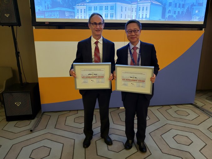
Jae K. Oh Profile Banner
Jae K. Oh Profile
Jae K. Oh

@JaeKOh2

7,206
Followers
228
Following
170
Media
342
Statuses

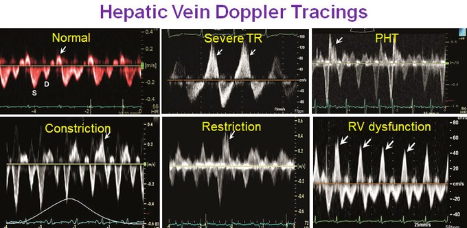
Cardiac Hemodynamics, Diastology, Pericardial Diseases, Valvular HD EVOID AS Trial & ECG/Echo AI

Mayo Clinic Rochester
Joined January 2014
Don't wanna be here? Send us removal request.
@JaeKOh2
Jae K. Oh
11 months
Chris Appleton and I have a sad news to share with the entire #Echo @ASE360 community.Liv Hatle, the legend, pioneer, and mentor to ALL of us passed away on 6/23. When we saw her a mo. ago, she was talking about her new book and calcific MS. Let us celebrate her amazing career!
Tweet media one
19
82
346
@JaeKOh2
Jae K. Oh
4 years
Thanks for sharing those fantastic IVC images and hepatic vein Doppler tracings. Hepatic vein Doppler taught by #LivHatle is critical for @aae @ASE360 echo assessing different underlying pathologies for congestion as well as volume status. Should be a part of comprehensive Echo.
Tweet media one
@ArgaizR
Eduardo R Argaiz
4 years
56
503
1K
2
139
298
@JaeKOh2
Jae K. Oh
3 years
@HeartDocSharon @MayoClinicCV @RigolinVera @JLindnerMD @WilliamZoghbi @ASE360 @StevenLesterMD Another critical Echo feature of constrction is preserved (>= 8cm/s) or ⬆️ mitral Medial Annulus e' velocity which is ⬇️ in ALL MYOCARDIAL DISEASES. When CP is mixed with myopathy, e' is lower than that in pure CP. Constriction until proven otherwise when e' is⬆️ in HF patient!
3
97
286
@JaeKOh2
Jae K. Oh
3 years
@HeartDocSharon @MayoClinicCV @RigolinVera @JLindnerMD @WilliamZoghbi @ASE360 @StevenLesterMD Thanks for tweeting this @HeartDocSharon who gave a great contrast talk @ASE360 . Here is real time animation #LivHatle and we @MayoClinicCV put together 20 years ago. The best demonstration of constriction hemodynamics. Do not miss constriction which is a CURABLE Diastolic HF.
12
108
267
@JaeKOh2
Jae K. Oh
11 months
#0 /7 Thanks @purviparwani for robust discussion on diastology which means "Dilation". I was asked to present my approach to Diastolic Function assessment at @ase360 " Just Relax: Diastolic Dysfunction" session. Let me share again and explain the 7 points that I emphasized.
Tweet media one
@purviparwani
Dr. Purvi Parwani
11 months
Stuck in echo lab today but loving Dr. @JaeKOh2 talk on diastology! Thank you @ASE360 for live stream! 1. E prime is the most important parameter 2. Systolic dysfunction doesn’t always mean diastolic dysfunction 2. Not all the diastolic parameters are required 3. Variability…
Tweet media one
Tweet media two
Tweet media three
Tweet media four
11
147
435
11
112
261
@JaeKOh2
Jae K. Oh
7 months
A 65 yo pt was referred to cardiac surgery for pericardiectomy with CT and cath (see below) consistent with constriction. Intraoperative @ase TEE was performed by an outstanding anesthesiologist. What do you think he said to the surgeon who is about to do sternotomy? @aae_echo
Tweet media one
Tweet media two
20
50
233
@JaeKOh2
Jae K. Oh
3 years
It is a myth that diastolic function is always abnormal in patients with systolic dysfunction. Young patients with reduced EF can have normal DF and filling pressure. Shown below was obtained from a 40 year old woman with LVEF of 25%. Filling pressure is normal. @jamil_tajik
Tweet media one
@iceman_ex
Segun Olusanya (He/Him) [email protected]
3 years
@apuxty @Wilkinsonjonny @NephroP @kyliebaker888 @cianmcdermott @susannaprice For me that’s a problem, and there are several actually: 1. Diagnosing anyone with systolic dysfunction with diastolic dysfunction is pointless as by definition the two go together 2. In ICU loading conditions constantly change so what looks like grade 1 can actually be grade 2
2
0
13
14
78
233
@JaeKOh2
Jae K. Oh
4 years
I was trying to emphasize that LV diastolic dysfunction cannot happen with normal myocardial relaxation. The best way to assess LV relaxation is mitral annulus e' velocity. So, medial or septal e' >=9 cm/sec indicates normal relaxation and diastolic function. @aae_echo @ASE360
@MonSangh
Monika Sanghavi MD, FACC
4 years
If septal e’>15, diastolic function is NORMAL - no additional assessment is needed. @JaeKOh2 #CVIECHO2020
Tweet media one
4
29
77
6
69
222
@JaeKOh2
Jae K. Oh
3 years
@argulian This HV Doppler shows increased inspiratory diastolic flow reversal cw high RV DP due to a myocardial disease. The last cycle was with inspiration since forward flow velocity increased. Diastolic reversal happens with expiration in constriction. @jamil_tajik Not TR! @EchoCases
Tweet media one
3
48
215
@JaeKOh2
Jae K. Oh
11 months
1/3. @EleidMack asked me to review Echo which was interpreted as normal. Looking at closely, Echo shows a subtle septal motion change. M-mode of LV would have shown septal motion change more clearly. Mitral inflow velocity showed >25% respiratory variation suggestive of CP.
Tweet media one
@EleidMack
Mack Eleid
11 months
0
1
8
7
60
204
@JaeKOh2
Jae K. Oh
3 years
1/2 Just received a box of chocolate from my pt who underwent pericardiectomy during COVID by @MayoClinicCVS after several yrs of HF elsewhere. Had numerous thoracenteses and liver evaluations including a biopsy. #Echo diagnosed CP using #Hatle and @MayoClinicCV dx criteria.
Tweet media one
5
44
196
@JaeKOh2
Jae K. Oh
3 years
Doppler, Color M mode, and strain from a patient with HCM. A good example of short IVRT and "L" wave indicating increased filling pressure. E velocity flow propagation velocity does not work in a small LV as in HCM. @jamil_tajik @ASE360 @aae_echo @KyleWKlarich @DavidWienerMD
Tweet media one
Tweet media two
Tweet media three
4
58
188
@JaeKOh2
Jae K. Oh
3 years
From @MayoClinicCV #hemody session. PV premature diastolic opening is related to increased RVEDP, but not necessarily increased mean RV diastolic pressure. (See 2 examples below) Same in the LV. @jamil_tajik @ASE360 @aae_echo @EchoCases
Tweet media one
2
34
156
@JaeKOh2
Jae K. Oh
5 years
This is an amazing prognostic data using diastolic exercise use echo 15 years after our initial feasibility study. Diastolic exercise echo is most helpful in patients with grade 1 diastolic dysfunction and exertional dyspnea. Essential for DX of HFpEF or diastic heart failure.
Tweet media one
@garvankane
Garvan Kane
5 years
Tweet media one
0
3
14
3
64
149
@JaeKOh2
Jae K. Oh
11 months
3/3. This is an educational case. Please review the Echo Diagnsotic Criteria for #constriction which works for A. FIB as well as in sinus rhythm.
Tweet media one
6
35
148
@JaeKOh2
Jae K. Oh
4 months
How embarrassing🥶 my mistake posting my photo👎 I was visiting my 95 yo mother and took few pictures. Thought nice to use one of them to my profile, but did not realize it would be posted. Well, my mom & I like to take this chance to wish everyone a Happy New Year of Dragon!
Tweet media one
Tweet media two
@EchoSoliman
Hatem Soliman, FRCP, FEACVI, FASE, FHEA
5 months
@JaeKOh2 Great to see you Dr Oh! Happy New Year 🎉😊
0
0
0
17
2
146
@JaeKOh2
Jae K. Oh
4 months
Exciting to start New Year 2024 with publication of our work on "Artificial Intelligence enabled ECG for diastolic function and filling pressure". Diastolic function by AI-ECG has a similar prognostic value as that of Echo. @drpaulfriedman @MayoClinicCV
5
49
142
@JaeKOh2
Jae K. Oh
5 months
Tweet media one
1
3
142
@JaeKOh2
Jae K. Oh
11 months
Most cardiologists were skeptical on her initial data on Doppler hemodynamics, but she was persistent to make Echo- Doppler as the current Hemodyanamic Gold-Standard ! We thank her for perseverance, brilliance, humbleness, and friendship. She loved her rugs, garden, & whiskey !
Tweet media one
Tweet media two
Tweet media three
Tweet media four
1
24
128
@JaeKOh2
Jae K. Oh
2 years
#LivHatle flew 26 hrs to energize the #ASE2022 . Her Doppler work that many doubted possible has made Echo as the clinical hemodynamic GOLD standard in valve, diastolic dysfunction, & pericardial diseases! We still need more education to help our pts by what she began in 1976.
@iamritu
Ritu Thamman MD
2 years
Tweet media one
0
10
51
1
14
122
@JaeKOh2
Jae K. Oh
3 years
1/2 I believe diastolic function assessment is the key to diagnosis of HFpEF. We need to show increased filling pressure at rest or with exercise to diagnose HFpEF. A promising new parameter is LA reservoir strain. It needs to be incorporated into the next diastic GL.
Tweet media one
@hahn_rt
RTHahnMD
3 years
The incomparable @JaeKOh2 speaking about Diastolic Function—so many NEW parameters to improve Sn/Sp!! You can still see the recorded ECHO/IMAGING New York State-of-the-Art 2021 by registering! @ASE360 @ACCinTouch @JudyHungMD @robertomlang @iamritu
1
45
125
2
44
120
@JaeKOh2
Jae K. Oh
3 years
Another variation of this theme is assessing AS in A Fib with different cycle lengths. #Nkomo @MayoClinicCV have the data that a single highest LVOT and AV velocities can reflect the severity of AS , better than averaging 3 or 5 cycles which is time consuming. See figure below.
Tweet media one
@AJamilTajik
Abdul Jamil Tajik
3 years
@RowseMD @RowseMD Post PVC LV contraction is potentiated + increased LVEDV, increased SV, result = increased gradient & PP in AS & decreased PP in HCM. May be LV in AS has novel response under GA in OR? @adnanalkhouli @JaeKOh2 @RickNishimura @fuadfehmi @mahali87
1
3
16
1
36
121
@JaeKOh2
Jae K. Oh
3 years
Beautiful study of ECP @NephroP Hepatic V Doppler confirms constriction. Few differences in Doppler between tamponade , ECP, and CP below. e' is not as high in tamponade and ECP as CP. Pure tamponade has gr 1 mitral inflow. Steroid if dialysis does not work. @jamil_tajik @ASE360
Tweet media one
@NephroP
NephroPOCUS
3 years
@VerwerftJan @JaeKOh2 @AllanLKleinMD1 @ArgaizR Potentially uremic. Better to start dialysis if pt agrees.
0
0
1
3
47
113
@JaeKOh2
Jae K. Oh
3 years
We @MayoClinicCV @garvankane tried to duplicate the Diamond-Forrester classification (NEJM 1976) using Echo hemodynamic assessment in cardiac ICU pts. Mortality is better overall, but Echo (E/e' and CI or SVI) classifies prognosis well. @jamil_tajik
2
37
113
@JaeKOh2
Jae K. Oh
6 years
I thank @MayoClinicCV , Liv Hatle, Jamil Tajik, Jim Seward and Pericardial Clinic/Echolab colleagues for receiving Lifetime Achievement Award from ESC Myo-Pericardial Disease working group at its meeting in Belgrade along with Allan Klein @ASE360 who was my fellow 30 years ago
Tweet media one
22
18
114
@JaeKOh2
Jae K. Oh
2 years
1/2 Pulmonary regurgitation velocity provides insights not only to right side hemodynamics, but also to left heart as shown in the figure (PR Doppler and LV/RV/RA). PR velocity decreases sharply at early diastole, then increases or plateaus at mid-diastole. Why?
3
26
114
@JaeKOh2
Jae K. Oh
11 months
#0 /7 Thanks @purviparwani for robust discussion on diastology which means "Dilation". I was asked to present my approach to Diastolic Function assessment at @ase360 " Just Relax: Diastolic Dysfunction" session. Let me share again and explain the 7 points that I emphasized.
Tweet media one
@purviparwani
Dr. Purvi Parwani
11 months
Stuck in echo lab today but loving Dr. @JaeKOh2 talk on diastology! Thank you @ASE360 for live stream! 1. E prime is the most important parameter 2. Systolic dysfunction doesn’t always mean diastolic dysfunction 2. Not all the diastolic parameters are required 3. Variability…
Tweet media one
Tweet media two
Tweet media three
Tweet media four
11
147
435
3
25
104
@JaeKOh2
Jae K. Oh
4 years
From @MayoClinicCV experience of using @ASE360 2016 guideline, we @garvankane feel that few modifications will make the guideline more practical and reliable for diastolic function and filling pressure assessment. Please try and share your experience.
Tweet media one
@drmalissawood
Malissa Wood
4 years
Superb discussion of brand new diastolic function update by 🌟⁦ @JaeKOh2 ⁩ ⁦ @JACCJournals ⁩ ⁦ @ASE360 #CVIECHO2020
Tweet media one
Tweet media two
1
10
25
5
49
104
@JaeKOh2
Jae K. Oh
3 years
@VivekKulkarniMD @ASE360 @HJarrett_MD @tiffchenMD @FSilvestryMD The figure shows mostly systolic flow reversal with inspiration. The configuration of systolic reversal is not typical for severe TR (late peaking). This is an early peaking inspiratory systolic flow reversal due to PHT or RV dysfunction. See below different HV Doppler signals.
Tweet media one
2
27
93
@JaeKOh2
Jae K. Oh
3 years
A good point @strain_rate Triangular TR is most commonly due to severe TR, but not always. Two examples below: (L) Increased flow to the RA from portal vein after TIPS and (R) Constriction. Unlike severe TR, TR jet is not dense. @jamil_tajik @ASE360
Tweet media one
Tweet media two
@strain_rate
Asbjørn Støylen 🇳🇴🇩🇰
3 years
Tweet media one
2
14
54
5
30
87
@JaeKOh2
Jae K. Oh
3 years
Respiratory variation of mitral E velocity becomes lessened when LA pressure is markedly elevated. The variation becomes unmasked if LA pressure or preload is reduced; Nitroglycerin as shown in theast tweet, or upright position as published below. @ASE360 @aae_echo
Tweet media one
@JaeKOh2
Jae K. Oh
3 years
Respiratory variation of mitral E vel. is a characteristic feature of CP as shown in the animation. But, we need to remember that about a 1/3 of CP patients have no or < 25% respiratory change. You can unmask the variation with certain maneuvers as below. @ASE360 @jamil_tajik
Tweet media one
Tweet media two
2
25
73
1
21
85
@JaeKOh2
Jae K. Oh
5 years
One of the most valuable diagnostic aspects of Echocardiogrpahy @ASE360 @echocardiac is its ability to assess cardiac hemodynamic conditions. Hope to share with you interesting and diagnostic Doppler tracings. Take a look at the CW Doppler recording from pulmonic valve.
Tweet media one
5
30
86
@JaeKOh2
Jae K. Oh
3 years
@garvankane @MayoClinicCV @AlsidawiMD & Dr. V. Nkomo published as below that the highest AS velocity when rhythm changed to AF was similar to peak AS velocity during NSR. This makes our AS assessment easier just using highest AV and LVOT V. Esquitin & @hahn_rt also published similar data in JASE 2019.
Tweet media one
3
33
84
@JaeKOh2
Jae K. Oh
5 months
Since there is a high interest in color M mode of hepatic vein diastolic reversal flow (red flow) with expiration in constrictive pericarditis, I like to share another one with corresponding PW Doppler in a pt with constriction and atrial fibrillation. @ecocardio_cl @AJamilTajik
Tweet media one
@JaeKOh2
Jae K. Oh
6 months
Yes. The color M mode beautifully demonstrates diastolic flow reversal with expiration which is diagnostic for constrictive pericarditis. You may want to share corresponding pulsed wave Doppler tracing.
4
15
82
4
24
85
@JaeKOh2
Jae K. Oh
3 years
We started the 4th day of #MayoEchoBR @garvankane with interesting cases. You will be interested in the following case and I want to know what you think. Exercise Dopper in 76 patient with exertional dyspnea and mitral annulus calcification. Also had ablation for AF. @ASE360
Tweet media one
11
20
83
@JaeKOh2
Jae K. Oh
4 years
A main point of the presentation was that medial e' velocity >=8 cm/s should raise a strong diagnostic concern for constrictive pericarditis in all patients with elevated jugular venous pressure and HF. e' is 5 cm/s or less in most,if not all, myocardial HFs. @ASE360 @aae_echo
@drankitjain
Ankit Jain MBBS,FASE,FASA,MBA.
4 years
Wonderful teaching by the one and only @JaeKOh2 @ASE360 #echohawaii . Hemodynamic s in Const Pericarditis and differentiating btw CP and RCM.
7
111
247
0
34
83
@JaeKOh2
Jae K. Oh
6 months
Yes. The color M mode beautifully demonstrates diastolic flow reversal with expiration which is diagnostic for constrictive pericarditis. You may want to share corresponding pulsed wave Doppler tracing.
@ecocardio_cl
Julián Vega Adauy
6 months
#EchoFIRST Hepatic vein M-COLOR mode is all you need! @JaeKOh2 would you agree?
Tweet media one
2
12
48
4
15
82
@JaeKOh2
Jae K. Oh
3 years
We just heard an amazing history of Hemodynamic #Echo from @jamil_tajik at @MayoClinicCV @Columbia MC #EchoImagingNY . We all are his students of Echo and cardiology. It was great to recognize contribution of late Mark Callahan who validated CW Doppler for AS. We all miss him.
Tweet media one
Tweet media two
2
16
78
@JaeKOh2
Jae K. Oh
5 years
Pearl's from @garvankane @MayoClinicCV #Echo BOARD Review on diastology. 1. E/e' >15 is specific but not sensitive for increased filling pressure except in MAC and LBBB. 2. L wave > 40 cm/ sec indicates usually grade 2 dysfunction. @ASE360
Tweet media one
Tweet media two
3
29
75
@JaeKOh2
Jae K. Oh
2 years
@CKFreeEcho @ASE360 @MDMankad @StevenNoyes @jstaffordmd @RobChamb87 @boegel_kelly @VLSorrellImages @HeartOTXHeartMD @rajdoc2005 @MorganC06473791 @a_l_bailey @EchoKen @echoguru The PV velocity is the famous "L" wave indicating delayed myocardial relaxation which reduces LV diastolic pressure resulting in an increased flow from PV to LA to LV. Systolic and diastolic PV velocities were called "J" and "K" , respectively. "L" comes after J and K.
4
19
74
@JaeKOh2
Jae K. Oh
3 years
Respiratory variation of mitral E vel. is a characteristic feature of CP as shown in the animation. But, we need to remember that about a 1/3 of CP patients have no or < 25% respiratory change. You can unmask the variation with certain maneuvers as below. @ASE360 @jamil_tajik
Tweet media one
Tweet media two
@JaeKOh2
Jae K. Oh
3 years
@HeartDocSharon @MayoClinicCV @RigolinVera @JLindnerMD @WilliamZoghbi @ASE360 @StevenLesterMD Thanks for tweeting this @HeartDocSharon who gave a great contrast talk @ASE360 . Here is real time animation #LivHatle and we @MayoClinicCV put together 20 years ago. The best demonstration of constriction hemodynamics. Do not miss constriction which is a CURABLE Diastolic HF.
12
108
267
2
25
73
@JaeKOh2
Jae K. Oh
7 months
This case illustrates the diagnostic importance of medial mitral annulus ( MA) e' velocity. TEE showed reduced motion of MA with e' of 4 cm/s which is diagnostic for cardiomyopathy. Surgery was cancelled. See video for annulus motion before & after typical pericardiectomy.
@MKaldas
Marco Kaldas
7 months
@JaeKOh2 @ASE @aae_echo He should tell him : This is a case of severe restrictive cardiomyopathy and the VSD influenced such cath hemodynamics
1
0
6
1
15
72
@JaeKOh2
Jae K. Oh
6 years
A great slide from @garvankane @MayoClinicCV at #MayoEchoBRC explaining the difference between normal (top) and restrictive filling with similar mitral inflow pattern. A major difference is the myocardial relaxation shown on 2D and mv annulus tissue Doppler. #AAE @ASEAN @ASE360
2
26
72
@JaeKOh2
Jae K. Oh
4 years
When I was learning Doppler, I saw a patient with acute severe AR with the similar findings. I thought the patient had restrictive CM and severe AR. #LivHatle smiled and I learned to differentiate restrictive CM from restrictive hemodynamics from the embarrassing experience.
@DrSergioBarros
Sergio Barros-Gomes, MD
4 years
What's the diagnosis of the M-mode across MV? Would you expect any abnormal murmur? How about the LV filling? Back to basics of #echofirst
Tweet media one
Tweet media two
7
31
70
1
20
68
@JaeKOh2
Jae K. Oh
2 years
@argulian The higher velocity is MR jet starting right after the closure of mitral inflow. The second jet with a dagger shape is probably from intracavitary gradient due to hyperdynamic heart. The CW cursor does not go thru the LVOT and the velocity peak is quite late. @AJamilTajik
1
6
69
@JaeKOh2
Jae K. Oh
1 year
We @MayoClinicCV just started a clinical trial to test whether DPP4 inhibitor can reduce progression of aortic stenosis after promising animal and retrospective human data . We began recruitment of subjects with mild to mod AS from 20 clinical sites.
Tweet media one
Tweet media two
Tweet media three
@MayoClinic
Mayo Clinic
1 year
Mayo Clinic is seeking adults with Aortic Stenosis (AS) to join a research study evaluating the effectiveness of a medication in delaying the progression of AS. Learn more at email RSTEVOID @mayo .edu or call 507-266-9861.
Tweet media one
2
6
9
0
12
68
@JaeKOh2
Jae K. Oh
4 years
The algorithm was proposed by @WilliamZoghbi group in Houston. I believe we can use the same algorithm for patients with primary pulmonary hypertension, LBBB , or even mitral prosthesis. @aae_echo @ASE360 E/e' may not be reliable in those situations.
@MonSangh
Monika Sanghavi MD, FACC
4 years
Proposed assessment of diastolic function in patients with mitral annular calcification. @JaeKOh2 #CVIECHO2020
Tweet media one
1
6
24
0
27
66
@JaeKOh2
Jae K. Oh
2 years
2/2 Simultaneous cath shows rapid rise of RVDP, then plateaus. LVDP also increases quickly, even higher than RVDP. The high LVDP is reflected to PADP to increase PR velocity at mid-diastole. Join #EchoAsia 2022 to hear about more interesting cases.
Tweet media one
0
19
62
@JaeKOh2
Jae K. Oh
3 years
Very good observation. She had mid-diastolic flow termed "L" wave. There 2 different L waves. One in normal subjects with bradycardia usually <40cm/sec as seen in her. This was first reported by G Keren et al. in Circulation 1986. Figure is from the paper. 10.1161/01.cir.74.1.36
Tweet media one
@prototypicaI
WC 🕙
3 years
@JaeKOh2 @jamil_tajik Looks like mid diastolic notching
0
0
0
1
11
59
@JaeKOh2
Jae K. Oh
3 years
Dr. Raul Espinosa is giving a wonderful talk on pericardial disease at #MayoEchoBR @garvankane . Just showed a characteristic LV strain pattern of constrictive pericarditis. Usually, inferior and lateral walls have reduced strain due to their tethering to the pericardium.
Tweet media one
0
21
60
@JaeKOh2
Jae K. Oh
11 months
2/3. In addition, mitral annulus e' was increased. Lateral e' was still slightly higher than medial e'. About 30% of CP patients do not have annulus reversus. In any pt with evidence of HF, mitral Medial e' > 8 cm/s strongly suggests CP. Hepatic vein Doppler would have been DX.
Tweet media one
2
8
60
@JaeKOh2
Jae K. Oh
3 years
The beginning of @MayoClinicCV Echo Lab and Doppler Echo. Dr. Liv Hatle was a great gift to us when she came to Mayo as a visiting professor. @jamil_tajik
Tweet media one
Tweet media two
Tweet media three
1
11
58
@JaeKOh2
Jae K. Oh
3 years
2/2 LA strain decreases as filling pressure increases. LAs < 25% usually indicates increased filling pressure as @robertomlang team initially showed. LAs is also helpful when there is a discrepancy between other diastolic parameters as shown below in a patient with MAC. Try it.
Tweet media one
Tweet media two
0
15
55
@JaeKOh2
Jae K. Oh
3 years
LVEF should 👍 in this pt since high mean gradient (>40 mmHg) is a good predictor. Very nice to obtain AV vel from SS fossa or notch! The highest AV vel is obtained from right para or SSF in 2/3. One caution! Vel from subclavian art stenosis can look like AS signal (Fig) @ASE360
Tweet media one
Tweet media two
@AJamilTajik
Abdul Jamil Tajik
3 years
@drEdgarFuentes @jorgeachv @jusalas77 @gutierrezjaikel @ASOCARCR @almasthela @MigueldeBoedo @RevEcocar @ImagenCardiaca @SISIACOficial @MAecocardio Beautiful images. Excellent demonstration of right supraclavicular fossa to record the highest velocity AS jet. RSCF, in selected patients does provide the unique “looking down the barrel” view of stenotic AV. Trust pt did well with AVR. @JaeKOh2 @dr_benoy_n_shah @BonitaEcho
0
1
10
0
8
55
@JaeKOh2
Jae K. Oh
1 year
It is very interesting to see the white demarcation line as @AJamilTajik has clearly shown in Fabry's disease. It is also interesting that endocardial portion of the postero-lateral segment is much thicker than that of anteroseptal segment with the whitish endocardial border.
@AJamilTajik
Abdul Jamil Tajik
1 year
Note the binary endocardial line on the apical long axis view, an important clue for Fabry's CM.
2
14
53
0
13
54
@JaeKOh2
Jae K. Oh
1 year
A beautiful CW Doppler tracing from @AJamilTajik a pioneer of #Echo . Typical tracing of mid ventricular obstruction, and it is interesting to see long IVRT (duration of the signal 3), and E velocity of about 50 cm/s indicating normal filling pressure in HCM.
@AJamilTajik
Abdul Jamil Tajik
1 year
Spectral Doppler demonstrated triple flow signals, resembling "Poseidon's Trident" Arrow: initiation of AR (onset of IVRT) 1 Aortic Flow during ejection 2 Mid ventricular obstruction 3 Flow during the IVRT from the apical aneurysm into LV sinus #Cardiotwitter #Echofirst
Tweet media one
11
34
130
1
14
56
@JaeKOh2
Jae K. Oh
3 years
Another L wave is seen in patients with high filling pressure and marked delay in myocardial relaxation which lowers LV pressure after its rise in early diastole. Usually > 40cm/sec as shown below along with corresponding PV (bottom) flow and MV color Mmode. @ASE360 @aae_echo
Tweet media one
1
11
53
@JaeKOh2
Jae K. Oh
4 years
The best method is contrast myocardial perfusion Echo. @HeartDocSharon Presence of perfusion in the area of apical abnormality is the key as shown. (L: apical perfusion, R: normal apex 2 mo later) This method will be ideal for COVID 19 situation. doi:10.1016/j.echo.2009.07.012
5
16
51
@JaeKOh2
Jae K. Oh
2 years
@AndreasGevaert @VerwerftJan @FH_Verbrugge @HerbotsLieven @nat_echo @echo_stepbystep @NephroP @ArgaizR @BarandiaranHF @JerremyWeerts @dr_benoy_n_shah E/e>15 is specific, but not sensitive for increased FP. In this pt, there is velocity below the baseline between Mitral E and A indicating ⬆️FP. IVRT appears to be shortened. However, TR velocity is normal. Need to know BP. LA strain appears to be ⬇️. PV be helpful. Overall ⬆️
4
13
52
@JaeKOh2
Jae K. Oh
6 years
Another name for this is "subepicardial aneurysm" Bunch et al. JASE 2003 16: 1318-21
@HeartToProve
Carlos El-Tallawi, MD “HeartToProve”
6 years
•False “pseudo” aneurysm: rupture w/ blood contained *outside* wall layers. (case below) •True aneurysm: no rupture; blood inside wall.
7
96
155
3
28
53
@JaeKOh2
Jae K. Oh
1 year
At the beginning of #EchoSOTA 2023 @ASE360 conference, Dr. Bijoy Khandheria was remembered and honored as a passionate #Echo educator and innovator by @WilliamZoghbi . His legacy will be our firm commitment to continue Echo education. @AJamilTajik @MayoClinicCV @MDMankad
Tweet media one
@AJamilTajik
Abdul Jamil Tajik
1 year
Gone too soon. May 11, 1956 - Feb 13, 2023. We mourn the sudden and untimely loss of a great person, Dr. Bijoy Khandheria. A top notch academic cardiologist, scholarly clinician, researcher, teacher, mentor and innovator.
Tweet media one
73
33
262
1
9
53
@JaeKOh2
Jae K. Oh
3 years
@jamil_tajik @mandeep_mayo @minhaskh @AllanLKleinMD1 @fuadfehmi @renujain19 @PatrycjaGalazka @Lakshmi14524052 @VLSorrellImages @ASE360 @mahali87 Effusive CP with mucomycosis. We need to treat her pericardial inflammation, preferably with steroid and colchicine, but challenging with fungal infection.If possible, we will get MRI and try to obtain pericardial fluid for ? fungal pericarditis. No annulus reversus in ECP.
Tweet media one
2
21
50
@JaeKOh2
Jae K. Oh
7 months
I like to invite you to ECHO ASIA 2023 to be held on Nov. 23 - 25 in Seoul, Korea. Outstanding faculty from @ASE360 @EACVIPresident and 15 @aae_echo member Societies. Share your best Echo case or research at the meeting.
Tweet media one
2
15
51
@JaeKOh2
Jae K. Oh
2 years
@echo_stepbystep @strain_rate @JGrapsa @AJamilTajik @rajdoc2005 @ASE360 @FabianKnebel @papadocardio @iamritu @DavidWienerMD @Ivan_Echocardio @alcantaramonica @alessia_gimelli @EACVIPresident @OAB1967 @Matte_Cameli @MAecocardio @fpmorcerf Apical sparing is a great addition to detect advanced #amyloid , but it can happen in other conditions. Attached apical sparing pattern was seen in a pt with a recent ❤transplant and cardiac biopsy was negative. LV strain as well as diastolic function became NL in 2 mo later.
Tweet media one
Tweet media two
4
9
50
@JaeKOh2
Jae K. Oh
3 years
@VLSorrellImages @ASE360 @CASEfromASE @JournalASEcho Doppler shows diastolic MR with 2 humps. There are 2 reasons for diastolic MR: high LV diastolic pressure (1) and atrial relaxation (2). Cath #WRMiranda shows them beautifully. Patient's HF was from severe AR causing high LV diastolic pressure. @jamil_tajik @aae_echo
Tweet media one
0
11
50
@JaeKOh2
Jae K. Oh
5 years
We @MayoClinicCV and @hahn_rt at Columbia began our first collaborative Echo NewYork cosponsored with @ASE360 . An amazing interest in Echo knowledge with more than 300 attendees. Will share interesting data with you next 3 days.
Tweet media one
1
10
49
@JaeKOh2
Jae K. Oh
5 years
Continuing the PV CW Doppler for hemodynamic assessment @JaeKOh2 Case #2 . Both tracings have a very short PHT, but from 2 different conditions. Let us @ASE360 #AAE figure out. Also join @garvankane @MayoClinicCV #echo Board Review next weekend in Rochester. No snow now.
Tweet media one
Tweet media two
4
18
50
@JaeKOh2
Jae K. Oh
5 years
This PV Doppler comes from a patient with acute severe mitral regurgitation causing premature closing of the PV due to a pulmonary arterial V wave. This was nicely demonstrated by Grose et al in Feb 1984 Circulation (right figure). @ASE360 #Echo #BoardReview @MayoClinicCV
Tweet media one
Tweet media two
4
23
49
@JaeKOh2
Jae K. Oh
4 years
We then decided to collect similar cases and published. All patients with severe acute AR had restrictive mitral inflow which returned to normal after AVR. Interestingly, early MV closure on M- mode was present only in 30%. @ASE360 @aae_echo
Tweet media one
1
13
46
@JaeKOh2
Jae K. Oh
3 years
Thank @HFpEF for tweeting this simple and practical clinical pearl! E/A ratio < 1 is the best diastolic filling pattern for patients with LV failure and worst pattern for patients with PAH.
@HFpEF
Sanjiv J. Shah, MD
3 years
#HFpEF pearl of the day: If a patient with HF symptoms and normal LVEF has E/A ratio < 1 and PA systolic pressure > 50 mmHg on echo, it’s more likely PAH, not HFpEF. Low E/A ratio is due to underfilled LV due to pulmonary vascular obstruction in setting of PAH.
4
56
295
1
8
47
@JaeKOh2
Jae K. Oh
3 years
The session was great. I also learned a lot from talk by @hvanspall . Very nice summary of drug trials in HFpEF.
Tweet media one
Tweet media two
@hvanspall
Harriette Van Spall, MD MPH
3 years
@DrEugeneYang @JaeKOh2 Don’t mess with the best!!
0
0
1
3
9
43
@JaeKOh2
Jae K. Oh
3 years
That RCT was performed by 4 Korean Medical Centers led by DH Kang, MD who also directed endocarditis RCT showing early surgery is beneficial. I believe we need to use more objective parameters such as strain, diastolic function & BNP to determine the timing of AVR. @jamil_tajik
@renujain19
Renu
3 years
@jamil_tajik @dr_benoy_n_shah @VerwerftJan @MayoClinicCV @JaeKOh2 @EleidMack @RickNishimura @pattypellikka @PanithayaC @adnanalkhouli @mahali87 @AnkurKalraMD @iamritu @mmamas1973 One very small RCT of peak >4.5, mean >50, early surgical intervention associated with better outcomes than “watchful waiting” in asymptomatic very severe AS. Positive stress tests were excluded.
Tweet media one
1
4
14
1
13
44
@JaeKOh2
Jae K. Oh
2 years
@kylalaraMD @MayoClinicCV @kylalaraMD Your question was perfect. If we understand basic concept of diastolic function, we can assess it by 1 or 2 parameters instead of 4. Mitral flow short DT and L wave in A fib as well as in NSR indicate high FP. High TR and E/e' confirm. See cath tracing. @AJamilTajik
Tweet media one
2
12
45
@JaeKOh2
Jae K. Oh
3 years
Speaking about TR, a dagger configuration of TR velocity usually indicates severe TR. Here is one exception, and hepatic vein Doppler suggests otherwise with flow reversals with expiration, not inspiration. Think about this, Master! @MayoClinic @aae_echo @ase Again, P=4V²
Tweet media one
Tweet media two
@AJamilTajik
Abdul Jamil Tajik
3 years
1/6 With the burgeoning intrest in tricuspid valve disease, propelled by the availability of multiple catheter based therapies for management of severe TR, increasingly we are seeing patients with marked IVC dilatation... #echofirst
Tweet media one
3
21
66
1
15
42
@JaeKOh2
Jae K. Oh
2 years
@iamritu @RUBraveEnough @Pooh_Velagapudi @UNMC_IM @nicoa002 @scahq @ASE360 EXCELLENT! Elevated e' = Eternity Medial e' > 9 cm/sec True Normal Medial e' 7 - 9 cm/ sec OK Medial e' < 6 cm/ sec SOB Yes, continue exercise to maintain e' higher than 7 as in the figure from an active 80 yo. # EchoHemodynamics
Tweet media one
2
16
42
@JaeKOh2
Jae K. Oh
6 months
#EchoAsia2023 10th anniversary of @aae_echo has attracted 860 participants and faculty from 15 Asian Pacific Societies. Among them are President of Korean, Thailand, and Singapore Echo Society or Chapter who were a fellow at @MayoClinicCV .
Tweet media one
@JaeKOh2
Jae K. Oh
6 months
The plenary session at #EchoAsia2023 was dedicated to #LivHatle . @MayoClinicCV #ChrisAppleton shared his experience as her first fellow. The session was followed by @aae_echo @ASE360 joint session where #BenEidem discussed CHD in the West and the East.
Tweet media one
Tweet media two
0
0
8
2
6
43
@JaeKOh2
Jae K. Oh
3 years
Dr. Ratnasari Padang gave a beautiful @MayoClinicCV #MayoEchoBR presentation on endocarditis including discussion on mycobacteria Chimaera. She even discussed Echo may not be in certain situation for the diagnosis of IE. @PanithayaC
Tweet media one
Tweet media two
2
10
40
@JaeKOh2
Jae K. Oh
3 years
This was published in EHJ HF.
Tweet media one
1
12
41
@JaeKOh2
Jae K. Oh
4 years
Don't be too concerned. Any guideline is just a recommendation for most but not all situations. Impossible to provide a diastolic function guideline which works for every situation. You are right for the hypovolemia situation. If well measured e' is normal, D function is normal.
Tweet media one
3
11
42
@JaeKOh2
Jae K. Oh
5 years
This diastology Webinar will take place at 11 am CST July 2nd in US. Case presentations to help diastolic function assessment. Relaxation is the key for diastole and we can assess that by 2D and Doppler mitral annulus motion. #EACVI @aae_echo @ASE360 @MayoClinicCV
@bogdan_popescu1
Bogdan A. Popescu
5 years
Join Otto Smiseth, Jae Oh and myself on Tuesday evening for a practical EACVI Webinar on diastolic function: cases, tips and tricks for daily practice. #EACVI @escardio @ThorEdvardsen @Cosyns @denisamuraru @DonalErwan @jumagne
1
16
30
0
21
40
@JaeKOh2
Jae K. Oh
3 years
@iamritu @MayoClinicCV @pattypellikka @ASE360 @JournalASEcho @amerjohri @DocStrom @LucySafi @hahn_rt @rajdoc2005 @NadeenFaza Thx @iamritu for tweeting @JournalASEcho paper by Saki @piyokokuma . Survival ⬆️after AVR in pts with moderate AS. She, Zhu & #Miranda also showed GLS can identify high risk in mod AS. Time for a trial to ✔AVR in moderate AS with NL EF. Based on❓parameters @jamil_tajik @hahn_rt
Tweet media one
Tweet media two
3
15
41
@JaeKOh2
Jae K. Oh
3 years
Jared showed PISA for mitral stenosis as well as mitral regurgitation. Move the color flow baseline shift towards the direction if the jet. So, opposite direction for MR and MS. @ASE360 @aae_echo
Tweet media one
@garvankane
Garvan Kane
3 years
Dr. Jared Bird discussing the power of Doppler Echocardiography. Key equations for the Boards. #MayoEchoBR @JaeKOh2 @MayoClinicCV
Tweet media one
2
21
53
2
13
39
@JaeKOh2
Jae K. Oh
2 years
1/2 The tracing came from a patient with severe RCM due to radiation, but a similar pattern can be seen in CP with more left side involvement, or mixed with myocardial disease. Here is another PR Doppler to analyze. @AJamilTajik @ASE360
Tweet media one
@azzamtarraf
Azzam Tarraf
2 years
@JaeKOh2 Restrictive?
0
0
0
2
13
40
@JaeKOh2
Jae K. Oh
1 year
#EACVI2023 was great. My best part was a visit to #Doppler pioneer/mentor #LivHatle at Calonge. We discussed how to assess calcific MS which is different from rheumatic MS. #BrilliantMind #LivHatle is completing an amazing book. She looked great! Hope for many more visits!
Tweet media one
Tweet media two
Tweet media three
Tweet media four
2
2
39
@JaeKOh2
Jae K. Oh
11 months
#6 /7. E/e' >=15 is specific for increased FP, but not sensitive. We need to consider high FP when E/e'>10 esp with TR> 2.8 m/sec. E/e' does not work well in mitral annulus calcification. Use mitral E/A, IVRT,PV, and LA strain. In HCM, E/e' >15 is sensitive, but not specific.
Tweet media one
4
8
37
@JaeKOh2
Jae K. Oh
3 years
Sure, but young individuals have sufficient diastolic reserve to maintain NL filling and pressure even in the setting of reduced LVEF, LVH, or myocardial infarct. E' may be lower than expected for the young, but can still be low normal to have NL filling. Not infrequent...
@AJamilTajik
Abdul Jamil Tajik
3 years
Jae, the slogan "diastole is the first to go" has stood the test of time for decades. Exceptions do exist like the example shown but the fundamental pathophysiologic Cascade is the truth & not myth.
3
11
73
2
4
37
@JaeKOh2
Jae K. Oh
4 years
Yes. Acute volume loading, high output heart-failure (obesity, shunt, hyperthyroidism, etc) and constrictive pericarditis. @aae_echo @ASE360 If JVP is elevated and e' is normal or elevated, should consider CP as your first differential.
@PerKarl14424229
Per Karlsson
4 years
@JaeKOh2 Can the filling pressures be elevated with a normal e'?
0
1
0
3
10
38
@JaeKOh2
Jae K. Oh
3 years
The last talk today at @MayoClinicCV #MayoEchoBR is being given by Joe Maalouf, MD about TEE from Abu Dhabi, UAE where is the chair of CV at Mayo Hospital SSMC. What a beautiful image of right pulmonary vein by TEE! Clockwise at 60 deg for RPV and countercw at 120 deg for LPV.
1
5
36
@JaeKOh2
Jae K. Oh
4 months
@thaiscoutinhoCV A great TEE image! I had a similar case in a 70 yo male who had TIA. He had low LAA emptying velocity and I thought thrombus was a possibility. With no change after 6 weeks of anticoagulation, a papilloma was robotically removed. A Happy New Year! @KyleWKlarich
@thaiscoutinhoCV
Thais Coutinho
5 months
Look what I found in a patient with recurrent stroke! #Echofirst
Tweet media one
4
8
32
0
10
36
@JaeKOh2
Jae K. Oh
5 years
I agree with Ritu. We will need more diagnostic and prognostic data of LA strain. Recommend LA strain as well as LV strain measurement in all patients to be familiar with these important functional parameters. @ASE360 @aae_echo
0
11
37
@JaeKOh2
Jae K. Oh
6 months
Join #ECHOASIA at the 10th anniversary of @aae_echo Asian Pacific Association of Echo along with 30th anniversary of Korean SE. Faculties from @ASE360 @EACVIPresident and 15 Asian Pacific Echo Societies. Nov 23 -25. Complimentary on line registration
@doc_jdam
Jose Donato Magno
6 months
ECHO ASIA + KSE! Nov. 23-25, 2023 Join us for yet another high-impact congress in echocardiography, featuring many of the leaders and experts from Asia and beyond! Register for free online participation @aae_echo #echofirst #EchoAsia @JaeKOh2
Tweet media one
Tweet media two
2
7
16
4
11
37
@JaeKOh2
Jae K. Oh
3 years
Thank @Ed_Alania for diagnostic images and HV Doppler of #constriction . ⬆️ HV diastolic flow reversal happens with lower forward flow velocity, c/w expiration. Apical 4 view shows ⬆️Annulus velocity. Diagnostic enough to proceed with pericardiectomy without hemodynamic cath.
@Ed_Alania
Edgardo Alania
3 years
👴 60yo + SOB #echofirst #whycmr ✅Abnormal septal motion. septal bounce ✅IVC enlargement ✅HV diastolic flow reversals with exp ✅Pericardium 4-5mm Constrictive Pericarditis @CyntiaMachain @ozlembilen2 @drahmedmohsen85 @SONECOM_AC @DrJayMohan @kinasa85 @JaeKOh2 @ImagenCardiaca
2
33
74
0
14
36
@JaeKOh2
Jae K. Oh
2 years
@onco_cardiology @MayoClinicCV What a timing for your tweet about the DVD on Dr. Hatle's talk on 30 years of Doppler hemodynamics, Juan! I am about to give a talk "Echo is clinical hemodynamic GOLD standard" at Euro Echo @EACVIPresident in an hour. Continuation of inspiration from Dr. Hatle and @AJamilTajik !
3
4
35
@JaeKOh2
Jae K. Oh
3 years
Yes, Dr. Tajik. P=4×V² has made Echocardiography a reliable hemodynamic as well as imaging tool. Here is an example of CW Doppler across the mitral valve in a young patient with Kono procedure, AR, and dyspnea. P=4×V² helped us to understand her hemodynamics. @aae_echo @ase
Tweet media one
@AJamilTajik
Abdul Jamil Tajik
3 years
@engineers_feed Nearly 7 decades later another simple but also profoundly impactful formula in Medicine & in particular Cardiology was described P = 4V². @JaeKOh2 @mahali87 @EchofirstB @ASE360
1
7
26
2
9
34
@JaeKOh2
Jae K. Oh
5 years
Echocardiography needs to be more quantitative since it is becoming the Gold Standard for hemodynamic assessment. The @ASE360 "Echo Formular Review Guide" should help hemodynamic quantification. Understanding the hemodynamic concept and good Echo techniques are essential.
@robertomlang
Roberto M Lang
5 years
Please check it out. New @ASE360 product " ECHO FORMULA REVIEW Guide. Excellent to improve your reports, learn and pass the NBE board #Echofirst exam. Beautiful illustrations that teach you how to acquire and measure. Buy it soon! @asemarketplace .com. See you in Portland
Tweet media one
4
50
134
0
8
32
@JaeKOh2
Jae K. Oh
3 years
The sunset is from one of @jamil_tajik our favorite places and the deer family is from our quiet backyard. @DrMarthaGulati @MarthaGrogan1
Tweet media one
Tweet media two
@AJamilTajik
Abdul Jamil Tajik
3 years
Challenge Accepted! Burning sky at dawn reflecting over the still Lake Michigan. @adnanalkhouli @JaeKOh2 @MarthaGrogan1 Post the quietest picture you have.
Tweet media one
4
0
16
1
0
33
@JaeKOh2
Jae K. Oh
3 years
When someone proves in a clinical study that the very complex formula to derive tau can help better than simple e' velocity, I will talk about that parameter. We are trying to be practical clinicians. Thanks for your passion for tau.
2
2
33
@JaeKOh2
Jae K. Oh
5 years
The second day of @MayoClinicCV #Echo board Review started with Dr. Nkomo discussing #aorticstenosis types. Here is an example of quadracuspid AS. @ASE360
0
5
30
@JaeKOh2
Jae K. Oh
3 years
2/2 After Echo Dx criteria for CP was established thanks to #Hatle @jamil_tajik , @MayoClinic pericardial cardiologists, Echolab and Cathlab, our CP Dx and pericardiectomy by excellent @MayoClinicCVS increased remarkably. Thank you all on behalf of our grateful 2,000 patients!
Tweet media one
0
4
31