
Natasha
@nat_echo
5,906
Followers
1,230
Following
1,346
Media
7,780
Statuses
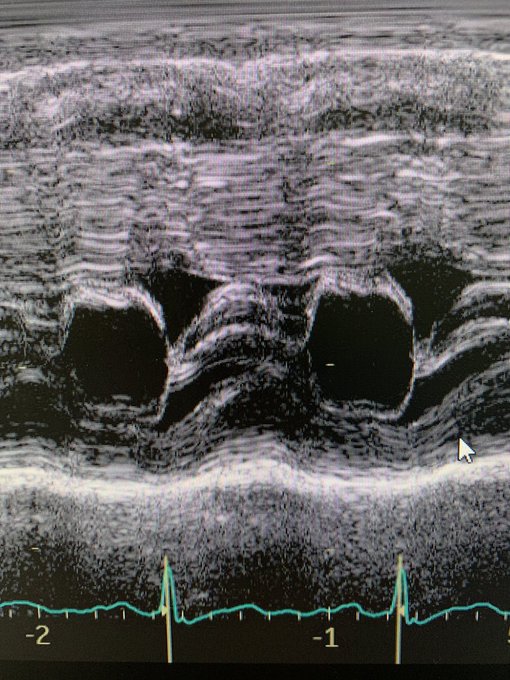
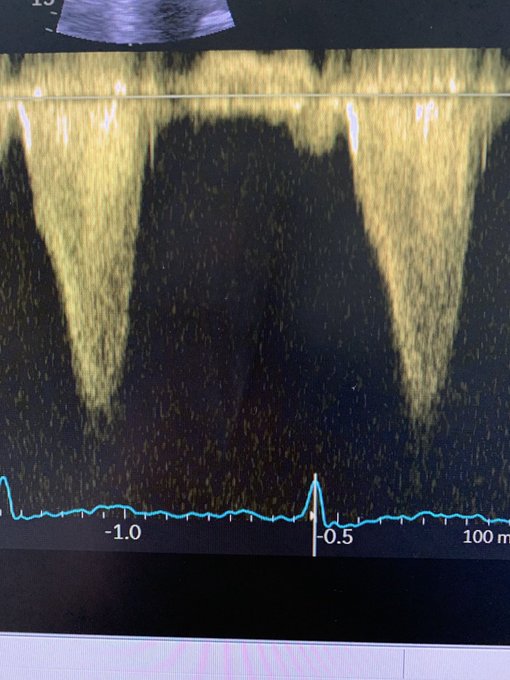
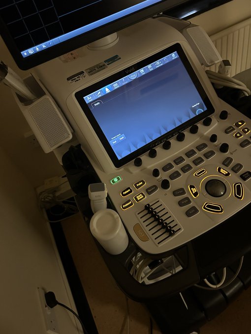
Cardiac Physiologist; “just like a fancy photographer” Enthusiastic about TOE & 4D echo. Vivid fan girl. Views my own. She/Her
England, United Kingdom
Joined April 2009
Don't wanna be here?
Send us removal request.