Dylan Burnette Profile Banner
Dylan Burnette Profile
Dylan Burnette

@MAG2ART

32,546
Followers
1,445
Following
1,493
Media
2,978
Statuses

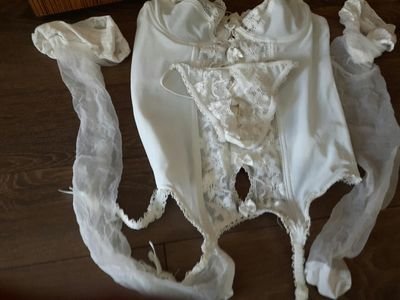
Cell biologist studying how a heart grows and dies. Associate Professor at Vanderbilt. Artist and fashion designer at . Married to @gillianhoo .

Nashville, TN
Joined January 2015
Don't wanna be here? Send us removal request.
Pinned Tweet
@MAG2ART
Dylan Burnette
5 months
DNA during cell death and cell division. #CellBiology #microscope
7
89
547
@MAG2ART
Dylan Burnette
6 years
THE "CITIZEN CANE"..... ......OF MICROSCOPY I'm teaching 1st-year grad students about cell migration tomorrow. This is the first movie they will see. An immune cell hunting down a bacterium among red blood cells. Movie credit: David Rogers. #CellBiology
168
2K
7K
@MAG2ART
Dylan Burnette
6 years
Cell 1: “I am going this way.” Cell 2: “No. You are coming with me.” #CellBiology #screenplay
59
1K
4K
@MAG2ART
Dylan Burnette
4 years
The Citizen Kane of Microscopy An immune cell (neutrophil) hunting down a bacterium among red blood cells. Filmed through a microscope by David Rogers. #CellBiology #Microscopy
82
875
3K
@MAG2ART
Dylan Burnette
3 years
Just a normal cell going through cell division.....WHAT? A dramatic cellular phenomenon? Let’s study that! #CellBiology #microscopy
52
591
3K
@MAG2ART
Dylan Burnette
1 year
Just a normal cell going through cell division…… Wait!!! What???!!! It was not so normal after all! I guess we have to study membrane blebs now…. The data said so.
48
419
3K
@MAG2ART
Dylan Burnette
2 years
A cancer cell with three nuclei and active mitochondria videoed through a microscope. @NikonSmallWorld
37
501
3K
@MAG2ART
Dylan Burnette
2 years
A cell going through cell division videoed through a microscope. Chromosomes are shown in pink. @NikonSmallWorld
29
521
3K
@MAG2ART
Dylan Burnette
5 years
DNA during cell death and cell division. #CellBiology #Microscopy
30
935
3K
@MAG2ART
Dylan Burnette
2 years
A cell going through cell division videoed through a microscope. #Science #CellBiology #Art
46
555
3K
@MAG2ART
Dylan Burnette
6 years
NEURONAL GROWTH CONES I spent years studying these little critters and I still do not understand them. Such is the burden that comes with choosing not to be a reductionist. Upside? Cells surprise me all the time. That has kept me coming back for 20 years. #CellBiology
49
585
2K
@MAG2ART
Dylan Burnette
5 years
NEURONAL GROWTH CONES I spent years studying these little critters and I still do not understand them. Such is the burden that comes with choosing not to be a reductionist. Upside? Cells surprise me all the time. That has kept me coming back for 20 years. #CellBiology
45
549
2K
@MAG2ART
Dylan Burnette
5 years
Mitochondria (green), microtubules (red) and actin filaments (blue) in a living cell as seen through a microscope. #CellBiology #MitoMonday
24
594
2K
@MAG2ART
Dylan Burnette
3 years
A cell going through cell division videoed through a microscope. #CellBiology #microscopy #SciArt
29
421
2K
@MAG2ART
Dylan Burnette
5 years
Cell 1: “I like going right.” Cell 2: “I like going left.” Cell 1: “I think I will go right forever.” Cell 2: “Nope. You’re coming with me.” Cell 1: “Where are we going?” Cell 2: “No idea but at least we’ll be together.”
27
462
2K
@MAG2ART
Dylan Burnette
5 years
Mitochondria (purple), DNA (yellow) and actin (blue) in an epithelial cell. @CellBiology @microscopy
30
503
2K
@MAG2ART
Dylan Burnette
3 years
Mitochondria (yellow) and microtubules (blue) in a cell videoed through a microscope. #CellBiology #microscopy #SciArt
32
393
2K
@MAG2ART
Dylan Burnette
6 years
Movie of a single cancer cell crawling around acquired through a microscope. Actin (purple); nucleus (yellow); Golgi (blue) #Cancer #CellBiology
45
655
2K
@MAG2ART
Dylan Burnette
2 years
Mitochondria and three nuclei in a cell videoed through a microscope. Only mitochondria videos until 30K (followers). #CellBiology
16
306
2K
@MAG2ART
Dylan Burnette
3 years
A cancer cell videoed through a microscope. It has three nuclei (blue). The mitochondria (yellow) and a cytoskeletal component (VASP;red) are also shown. #CellBiology #microscopy #SciArt
30
416
2K
@MAG2ART
Dylan Burnette
4 years
DNA (green) and mitochondria (purple) in a cancer cell as seen through a microscope. #CellBiology #microscopy #Melanoma
21
368
2K
@MAG2ART
Dylan Burnette
5 years
FULL OF ENERGY Mitochondria (purple) create much of the chemical energy for the cell. And when compared to the slow moving nucleus (green), mitochondria seem hyperactive. Full of energy indeed. #MitoMonday #CellBiology
31
580
2K
@MAG2ART
Dylan Burnette
1 year
Dying melanoma cancer cells videoed through a microscope. #CellBiology
38
233
2K
@MAG2ART
Dylan Burnette
5 years
Mitochondria (blue) and DNA (green) in a melanoma cell as seen through a microscope. #MitoMonday #CellBiology
25
489
2K
@MAG2ART
Dylan Burnette
1 year
Cells pretending to be galaxies. #CellBiology
Tweet media one
16
150
2K
@MAG2ART
Dylan Burnette
5 years
The powerhouses of our cells, mitochondria, moving around the nucleus. It reminds me of a toddler interacting with a patient golden retriever. #CellBiology #microscopy
12
419
2K
@MAG2ART
Dylan Burnette
5 years
Mitochondria (green), microtubules (red) and actin filaments (blue) in a living cell imaged through a microscope. #CellBiology #MitoMonday
18
475
2K
@MAG2ART
Dylan Burnette
3 years
A cell going through cell division photographed through a microscope. #CellBiology #microscopy #SciArt
Tweet media one
21
264
2K
@MAG2ART
Dylan Burnette
3 years
This is the first video of cell division captured in the Burnette lab. The DNA--starting in condensed chromosomes--and DIC are shown. Little did we know then that we would become obsessed with membrane blebs; apparent in this video during cytokinesis. #CellBiology #microscopy
28
382
2K
@MAG2ART
Dylan Burnette
3 years
Happy Valentine's Day! Here is a single heart muscle cell (cardiac myocyte)--which itself is shaped like a little heart--as seen through a microscope. #cardiotwitter #CellBiology #SciArt
18
408
2K
@MAG2ART
Dylan Burnette
5 years
Mitochondria (green) in a cancer cell with three nuclei (purple). #CellBiology #MitoMonday #microscopy
20
369
2K
@MAG2ART
Dylan Burnette
2 years
A dividing cell photographed through a microscope. #Science #Biology #CellBiology #Art
Tweet media one
11
253
2K
@MAG2ART
Dylan Burnette
4 years
DNA (yellow), actin filaments (blue), and the mighty mitochondria (purple) as seen through a microscope. #CellBiology #microscopy
28
327
2K
@MAG2ART
Dylan Burnette
5 years
The cell’s nucleus (blue), mitochondria (yellow), and focal adhesions (purple). #CellBiology #Microscopy
25
371
2K
@MAG2ART
Dylan Burnette
2 years
Neuronal growth cones videoed through a DIC microscope. #Science #Biology #CellBiology
20
183
2K
@MAG2ART
Dylan Burnette
4 years
This is a single cell as seen through a microscope. It shows the movement of mitochondria (green), microtubules (red) and actin filaments (blue). #CellBiology #Microscopy
25
370
2K
@MAG2ART
Dylan Burnette
4 years
The Citizen Kane of Cell Division Walther Flemming published this time-lapse of a dividing cell in 1882. He drew the data by hand while looking in his microscope. #CellBiology #microscopy #SciArt
16
303
2K
@MAG2ART
Dylan Burnette
3 years
Neuronal growth cones photographed using a microscope. #CellBiology #microscopy #SciArt
Tweet media one
12
218
2K
@MAG2ART
Dylan Burnette
4 years
This movie shows the powerhouses of the cell, mitochondria, and DNA within a single cancer cell with three nuclei as seen through a microscope. #CellBiology #Microscopy
20
332
2K
@MAG2ART
Dylan Burnette
4 years
The actin filament cytoskeleton in a single cell photographed through a microscope. #CellBiology #microscopy #SciArt
Tweet media one
24
234
2K
@MAG2ART
Dylan Burnette
3 years
A cell going through cell division photographed through a microscope. #CellBiology #microscopy #SciArt
Tweet media one
15
256
1K
@MAG2ART
Dylan Burnette
4 years
This is a single epithelial cell as seen through a microscope. The nucleus (blue), mitochondria (yellow) and the actin filament cytoskeleton (magenta) are shown. #CellBiology #microscopy
33
346
1K
@MAG2ART
Dylan Burnette
5 years
The "Citizen Kane" of Microscopy I am teaching first-year graduate students about cell motility tomorrow. This is the first movie they will see; a classic recording by David Rogers of a neutrophil chasing down a bacterium. #CellBiology #Microscopy
35
317
1K
@MAG2ART
Dylan Burnette
5 years
DNA during cell death and cell division. #CellBiology #microscopy
12
402
1K
@MAG2ART
Dylan Burnette
3 years
A dividing cell photographed through a microscope. Actin and myosin II filaments are shown. #CellBiology #Science #SciArt
Tweet media one
10
210
1K
@MAG2ART
Dylan Burnette
2 years
Three cells going through cell division videoed through a microscope. Left to right: Control, Myosin IIA depletion, and Myosin IIB depletion. This data screamed, "Study membrane blebs!" So we did.... #CellBiology #Biology #Science
16
240
1K
@MAG2ART
Dylan Burnette
1 year
A cell videoed through a microscope. DNA (cyan), mitochondria (yellow), and VASP (magenta) are shown. #CellBiology
16
239
1K
@MAG2ART
Dylan Burnette
4 years
Mitochondria (green), microtubules (red) and actin filaments (blue) in a living cell as seen through a microscope. #CellBiology
13
337
1K
@MAG2ART
Dylan Burnette
5 years
THE CELL’S POWERHOUSE Mitochondria (yellow) make much of the chemical energy that fuels the life of a cell. They also move rapidly as seen in this cancer cell compared to the relatively slow moving nucleus (blue) and cellular adhesions (purple). #MitoMonday #CellBiology
21
404
1K
@MAG2ART
Dylan Burnette
3 years
A single heart muscle cell (cardiac myocyte) videoed through the microscope. The spaghetti looking organelles are the powerhouses of the cell, mitochondria. Those white dots? You tell me... #CellBiology #cardiotwitter #microscopy
61
218
1K
@MAG2ART
Dylan Burnette
5 years
Some of my favorite cell-shape changes happen during cell division. #CellBiology
14
307
1K
@MAG2ART
Dylan Burnette
5 years
THE CELL’S POWERHOUSE Mitochondria (yellow) make much of the chemical energy that fuels the life of a cell. They also move rapidly as seen in this cancer cell compared to the relatively slow moving nucleus (blue) and cellular adhesions (purple). #MitoMonday #CellBiology
17
401
1K
@MAG2ART
Dylan Burnette
6 years
FULL OF ENERGY Mitochondria (purple) create much of the chemical energy for the cell. And when compared to the slow moving nucleus (green), mitochondria seem hyperactive. Full of energy indeed. #MitoMonday #CellBiology
22
522
1K
@MAG2ART
Dylan Burnette
3 years
A mitochondrion dancing on microtubules in a living cell as seen through a microscope. #CellBiology #microscopy #SciArt
29
216
1K
@MAG2ART
Dylan Burnette
2 years
A cell videoed through a microscope. DNA in the nucleus (green) and the powerhouses/overlords of the cell, mitochondria (purple), are shown. If your biology teacher taught you that mitochondria look like little beans, you now know better. #Science #Biology #CellBiology #Art
33
258
1K
@MAG2ART
Dylan Burnette
5 years
THE "CITIZEN CANE"..... ......OF MICROSCOPY An immune cell hunting down a bacterium among red blood cells. A picture may be worth a thousand words, but a movie can contain exponential insights. Movie credit: David Rogers ( @VanderbiltU , 1950's). @VUBasicSciences
17
444
1K
@MAG2ART
Dylan Burnette
6 years
POWERHOUSE AND HARD DRIVE Watching the powerhouse of the cell - mitochondria (yellow) - can be as mesmerizing as watching a camp fire. DNA (blue) is not as fun at this timescale. Luckily there are people who can image DNA better than me. #MitoMonday #CellBiology
18
408
1K
@MAG2ART
Dylan Burnette
2 years
A cell videoed through a microscope. The powerhouses of the cell, mitochondria (purple), and DNA in the nucleus (green) are shown. #CellBiology
32
264
1K
@MAG2ART
Dylan Burnette
3 years
The Citizen Kane of Cell Division Walther Flemming published this time-lapse of a dividing cell in 1882. He drew the data by hand while looking in his microscope. #CellBiology #microscopy #SciArt
10
264
1K
@MAG2ART
Dylan Burnette
3 years
The powerhouses/overlords of the cell, mitochondria (yellow), and DNA in the nucleus (blue) videoed through a microscope. #CellBiology #microscopy #SciArt
19
258
1K
@MAG2ART
Dylan Burnette
2 years
A mother cell dividing into two daughter cells photographed through a microscope. @NikonSmallWorld
Tweet media one
17
215
1K
@MAG2ART
Dylan Burnette
5 years
POWERHOUSE AND HARD DRIVE Watching the powerhouse of the cell - mitochondria (yellow) - can be as mesmerizing as watching a camp fire. DNA is blue. #CellBiology
18
358
1K
@MAG2ART
Dylan Burnette
5 years
The powerhouses of the cell, mitochondria, doing what they do. #MitoMonday #CellBiology
23
315
1K
@MAG2ART
Dylan Burnette
3 years
The powerhouse of our cells, mitochondria (green), and the genetic information in our cells, DNA (purple), videoed in a cancer cell with three nuclei using a microscope. #CellBiology #microscopy
19
271
1K
@MAG2ART
Dylan Burnette
6 years
CANCER CELL ON THE MOVE viewed through a microscope #Cancer #CellBiology
32
385
1K
@MAG2ART
Dylan Burnette
1 year
The leading edge of a cell videoed through a microscope. Actin filaments and adhesions are shown. #CellBiology
18
170
1K
@MAG2ART
Dylan Burnette
2 years
A single cell photographed through a microscope. DNA in the nucleus, the Golgi apparatus, and actin filaments are shown. The cell is sitting on a circular substrate. #CellColorBlast #CellBiology #Biology #Science #Art
16
248
1K
@MAG2ART
Dylan Burnette
2 years
A colony of stem cells photographed through a microscope by @nilaytaneja . #Science #Nature #CellBiology #Art
Tweet media one
21
200
1K
@MAG2ART
Dylan Burnette
4 years
This photograph of a cell kind of looks like a person. #CellBiology #microscopy
Tweet media one
36
135
1K
@MAG2ART
Dylan Burnette
4 years
A cancer cell with three nuclei (purple) and mitochondria (green) as seen through a microscope. #melanoma #CellBiology #microscopy
24
305
1K
@MAG2ART
Dylan Burnette
4 years
Neuronal growth cones as seen through a microscope. Movie shows a time lapse of the extensions from a cultured bag cell neuron isolated from Aplysia californica (i.e., a giant sea slug). #CellBiology #microscopy
14
232
1K
@MAG2ART
Dylan Burnette
2 years
A cell videoed through a microscope. DNA in the nucleus and the powerhouses/overlords of the cell, mitochondria, are shown. #Science #Nature #CellBiology #Art
14
228
1K
@MAG2ART
Dylan Burnette
5 years
A crawling cancer cell as seen through a microscope. #CellBiology #Melanoma
16
299
1K
@MAG2ART
Dylan Burnette
3 years
A cell videoed through a microscope. DNA in the nucleus (blue), mitochondria (yellow), and the cytoskeletal protein VASP (purple) are shown. #CellBiology #microscopy #SciArt
29
262
1K
@MAG2ART
Dylan Burnette
2 years
Mitochondria (top) and microtubules (bottom) in the same region of a cell videoed through a microscope. #Science #Biology #CellBiology #Art
12
200
1K
@MAG2ART
Dylan Burnette
3 years
Neuronal growth cones videoed through a microscope. They are quite active little critters; if they are happy. #CellBiology #microscopy #SciArt
17
220
1K
@MAG2ART
Dylan Burnette
4 years
A cancer cell dividing a seen through a microscope. #CellBiology #microscopy
20
330
1K
@MAG2ART
Dylan Burnette
3 years
Who is watching mitochondria change shape inside of a live cell? #CellBiology #microscopy #SciArt
17
175
1K
@MAG2ART
Dylan Burnette
5 years
Movie of mitochondria (green), DNA (blue) and actin (red) in an epithelial cell acquired with a spinning disk confocal microscope. #MitoMonday #CellBiology
22
341
1K
@MAG2ART
Dylan Burnette
5 years
Neuronal growth cones integrating information from their environment. 1- “Hey ya’ll. Are we on glass.” 2- “Yup” 3- “Yup” 4- “Yup” 1- :-/ A #CellBiology story.
14
282
1K
@MAG2ART
Dylan Burnette
4 years
A crawling cancer cell as seen through a microscope. The nucleus (yellow), Golgi apparatus (blue) and actin cytoskeleton (red) are shown. #CellBiology #Microscopy #Melanoma
10
255
1K
@MAG2ART
Dylan Burnette
3 years
A cell photographed through a microscope. #CellBiology #microscopy #SciArt
Tweet media one
11
152
1K
@MAG2ART
Dylan Burnette
3 years
DNA (green) and mitochondria videoed in a single cancer cell using a microscope. Mitochondria play multiple roles inside of a cell (e.g., powerplant, judge, and executioner); all while impersonating worms at a dance party. #CellBiology #microscopy #SciArt
17
227
1K
@MAG2ART
Dylan Burnette
3 years
A dividing cancer cell photographed with a microscope. Actin filaments, microtubules and DNA are shown. #CellBiology #microscopy #SciArt
16
180
1K
@MAG2ART
Dylan Burnette
5 years
POWERHOUSE AND HARD DRIVE Watching the powerhouse of the cell - mitochondria (yellow) - can be as mesmerizing as watching a camp fire. DNA (blue) is not as fun at this timescale. Luckily there are people who can image DNA better than I can. #MitoMonday #CellBiology
27
318
1K
@MAG2ART
Dylan Burnette
2 years
A cell videoed through a microscope. DNA in the nucleus (red) the powerhouses/overlords of the cell, mitochondria (cyan), and actin filaments (gray) are shown. #Science #Biology #CellBiology #Art
17
175
1K
@MAG2ART
Dylan Burnette
5 years
For those who like membrane blebs during cell division.
21
213
1K
@MAG2ART
Dylan Burnette
4 years
The first movie of cell division made in the Burnette lab; captured by ⁦ @AidanFenix ⁩. #CellBiology #microscopy
8
224
1K
@MAG2ART
Dylan Burnette
2 years
A cell photographed through a microscope. #CellBiology
11
199
1K
@MAG2ART
Dylan Burnette
5 years
Who is watching mitochondria change shape? #CellBiology #Microscopy
12
221
1K
@MAG2ART
Dylan Burnette
5 years
The powerhouses of our cells, mitochondria, are not all bean-shaped as your high school text book would you have you believe. As soon as you define their shape, they change it. Mitochondria are not into labels. #MitoMonday #CellBiology #Microscopy
19
328
1K
@MAG2ART
Dylan Burnette
4 years
A TALE OF TWO CELLS One dies; One divides. DNA is shown. It's not a long tale as it turns out. #CellBiology #Microscopy
12
252
1K
@MAG2ART
Dylan Burnette
2 years
Dying melanoma cancer cells videoed through a microscope. I find the moment when they finally "pop" quite satisfying. Notice the "apoptotic bodies" that were released from the cells scattered around the dish. #ScienceTermOfTheWeek . #Science #Biology #CellBiology #Art
12
206
1K
@MAG2ART
Dylan Burnette
4 years
A cancer cell as seen through a microscope. The three nuclei in this cell (blue), mitochondria (yellow) and the cytoskeleton (red) are shown. #CellBiology Should a cell have three nuclei? No. Explanations #NSGC20 ? Genomic instability? Cytokinesis failure?
32
213
1K
@MAG2ART
Dylan Burnette
3 years
Tweet media one
12
164
1K
@MAG2ART
Dylan Burnette
4 years
The is a single cell as seen through a microscope. Actin filaments (purple), mitochondria (blue) the nucleus (orange) are shown. The excitation light caused phototoxicity. Does anyone know how I figure that out? #CellBiology #Microscopy #SciArt
37
173
1K
@MAG2ART
Dylan Burnette
10 months
A cell photographed through a microscope. DNA in the nucleus, Golgi apparatus, and actin filaments are shown. #CellBiology
10
202
1K
@MAG2ART
Dylan Burnette
11 months
A cell videoed through a DIC microscope. The worm-like organelles are mitochondria and the circle at the bottom is the nucleus. #CellBiology
15
187
1K
@MAG2ART
Dylan Burnette
2 years
A cell videoed through a microscope. DNA in the nucleus (blue), the powerhouses/overlords of the cell, mitochondria (yellow), and actin filaments (purple) are shown. #Science #Biology #CellBiology #Art
16
202
997
@MAG2ART
Dylan Burnette
2 years
The powerhouses/overlords of the cell, mitochondria, videoed through a microscope. Yeah, I know. Not as many bean-like shapes as you were expecting? Your textbooks lied to you. #Science #Biology #CellBiology #Art
29
216
1K