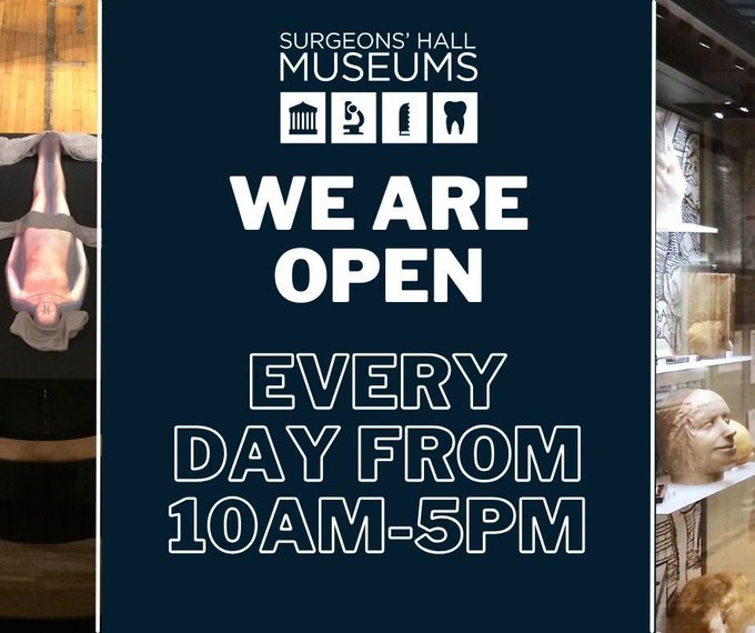
Home to the largest & most historic collection of surgical pathology in the world.
Open 10am-5pm, 7 days a week.
For Library and Archive follow
@RCSEDArchive
The Surgeons' Hall Riot happened
#OTD
in 1870. The Edinburgh 7 arrived at Surgeons' Hall to sit their anatomy exam & were met by a crowd of male students & several hundred onlookers who opposed of women studying medicine. Not to be deterred the women went on to sit their exam.
Surgeons use ligatures to tie off blood vessels during surgery to stop them from bleeding. These ligatures date from the late 19thc. & are made from horse hair. The fineness of the hair made it useful for both sutures & ligatures. The hair was sterilized & sold in rolls of 100.
The Surgeons' Hall Riot happened
#OTD
in 1870. The Edinburgh 7 arrived at Surgeons' Hall to sit their anatomy exam & were met by a crowd of male students & several hundred onlookers who opposed of women studying medicine. Not to be deterred the women went on to sit their exam.
This huge kidney stone has taken the shape of the pelvis and calyces of the kidney from whence it came, effectively forming a cast of the spaces inside the kidney. Kidney stones like this are known as staghorn calculi.
This length of insulated TV cable was removed from the bladder of a 23 year old man. It had become encrusted with phosphate and had coiled to the anatomical curves of the bladder.
The Surgeons' Hall Riot happened
#OTD
in 1870. The Edinburgh 7 arrived at Surgeons' Hall to sit their anatomy exam & were met by a crowd of male students & several hundred onlookers who opposed of women studying medicine. Not to be deterred the women went on to sit their exam.
The Surgeons' Hall Riot happened
#OTD
in 1870. The Edinburgh 7 arrived at Surgeons' Hall to sit their anatomy exam & were met by a crowd of male students & several hundred onlookers who opposed of women studying medicine. Not to be deterred the women went on to sit their exam.
A resin cast of the renal circulation of the left kidney. Different coloured resin has been used to distinguish veins (red), arteries (blue) & renal pelvis (yellow). Once the resin had been injected and set, the kidney tissues were then dissolved in 50% hydrochloric acid.
This skull shows the effects of Syphilis. The outer and inner tables of the skull have been eroded, while the diploe – the layer in between – has become thickened.
This huge kidney stone has taken the shape of the pelvis and calyces of the kidney from whence it came, effectively forming a cast of the spaces inside the kidney. Kidney stones like this are known as staghorn calculi.
The Surgeons' Hall Riot happened
#OTD
in 1870. The Edinburgh 7 arrived at Surgeons' Hall to sit their anatomy exam & were met by a crowd of male students & several hundred onlookers who opposed of women studying medicine. Not to be deterred the women went on to sit their exam.
The arteries of this hand have been preserved by being injected with wax. This specimen was prepared by surgeon Robert Liston. Injecting vasculature with wax (and more recently plastic) and then dissolving the surrounding tissue is called corrosion casting.
These casts show a woman with a large fibroma before and after surgery. Robert Liston removed the maxilla, upper jaw and the tumour in 1837. The woman survived this very risky procedure.
A glass bottle of sterlised catgut ligature from the 19th century. Only a few centimeters of the catgut still remain on one of the three bobbins inside the bottle. While called catgut, the sutures are usually made from sheep or goat intestines.
This upper end of the right femur is from a solider wounded by a musket ball at the Battle of Waterloo,
#OTD
in 1815. The impact of the musket ball has created a deep cavity in the neck of the femur in which the ball is almost completely embedded.
Dr. Elsie Inglis, founder of the Scottish Women's Hospitals, died
#OTD
in 1917. During WWI Inglis offered the British War Office a medical unit staffed entirely by women. Her offer was rejected & Elsie was told to 'go home and sit still.’ Her offer was accepted by the French Gov.
This knuckle-duster belonged to dentist Frederick Smith. Smith was a dentist in Chesterfield. He carried this knuckle-duster as protection from "highwaymen" on his weekly horse drawn cab journey to his practice in Alfreton.
Surgeons use ligatures to tie off blood vessels during surgery to stop them from bleeding. These ligatures date from the late 19thc & are made from horse hair. The fineness of the hair made it useful for both sutures and ligatures. The hair was sterilised and sold in rolls of 100
A skull showing the effects of Syphilis. The outer and inner tables of the skull have been eroded, while the diploe – the layer in between – has become thickened.
These sutures are made from kangaroo tendon. This glass phial is from the early 20th century and the sutures are in a carbolized glycerine. Kangaroo tendon is incredibly strong making the sutures suitable for deep tissue procedures.
This knuckle-duster was carried by Frederick Smith. Smith was a dentist in Chesterfield from 1862 to 1912. He carried this to protect himself against highwaymen on the weekly journey by horse drawn cab to and from his branch practice at Alfreton.
Upper end of the right femur of a solider wounded by a musket ball at the Battle of Waterloo, 18th June 1815. The impact of the musket ball has created a deep cavity in the neck of the femur in which the ball is almost completely embedded.
This knuckle-duster belonged to dentist Frederick Smith. Smith was a dentist in Chesterfield. He carried this knuckle-duster as protection from "highwaymen" on his weekly horse drawn cab journey to his practice in Alfreton.
Chain saws like this were used when access to the bone to be cut was too limited for a hand saw. One end of the chain would be detached from the handle and hooked around the bone using a carrier needle. This chain saw dates from the mid-19th century.
The upper end of the right femur of a solider wounded by a musket ball at the Battle of Waterloo, 18th June 1815.
The impact of the musket ball has created a deep cavity in the neck of the femur in which the ball is almost completely embedded.
A metal pill box from 1847. The compartments are marked with the name of the medication such as "grey powders". The box still contains some pills.
#histmed
Surgeons use ligatures to tie off blood vessels during surgery to stop them from bleeding. These ligatures date from the late 19thC & are made from horse hair. The fineness of the hair made it useful for both sutures & ligatures. The hair was sterilized & sold in rolls of 100.
This metal pill box dates from 1847. The compartments are marked with the name of the medication such as "grey powders". The box still contains some pills.
This wax model is on loan to us from the Gordon Museum of Pathology
@kingsmedicine
. It was created by Joseph Towne in the 1800s. It shows a cadaver with the brain and right lung removed . Towne's models were depicted exactly as they were seen in the dissection room and are [1/2]
Conditions in operating theatres prior to antisepsis were appalling. Most people were admitted in their ordinary clothes and surgeons operated in a suit and frock coat. This coat belonged to Joseph Lister and similar coats would be worn by a surgeon when operating.
Some of the instruments in this postmortem set from the 1850s include a skull saw, costotome (hammer), sprung tweezers, bowel scissors, chisel and a rachitome chisel. A blow pipe and more needles would have originally been included in the set but have sadly been lost over time.
A glass bottle of sterlised catgut ligature from the 19th century. Only a few centimeters of the catgut still remain on one of the three bobbins inside the bottle. While called catgut, the sutures are usually made from sheep or goat intestines.
An x-ray of Joseph Lister's hand.
This was made in November 1896 at King’s College Hospital, London within 18 months of Röntgen’s discovery of X-rays. The exposure time was said to be two minutes.
These little tools are apple scoops. They date from the 1800's and are made of sheep bone. They were common in apple-growing districts such as Somerset, Devonshire and Herefordshire to allow individuals with few or no teeth to enjoy apples.
Circle of Willis presented about 1897. This is a network of arteries that supplies blood to the brain. Arthur Conan Doyle mentions this in a poem of his about sitting an anatomy exam while he was a student.
This wooden mouth gag from the nineteenth century, would have been inserted in a gap between the teeth & screwed so that the patient opened their mouth. With the gag in place the mouth stayed open, this was particularly useful if the patient had a condition such as lockjaw.
#OTD
in 1834, Isabel Thorne was born. Thorne was part of the 'Edinburgh 7', the first group of women to study at any British University. Despite passing all their exams the women weren't allowed to graduate. You can join us in celebrating the 7 next month:
This wooden mouth gag (or prop) from the 19th century, would have been inserted in a gap between the teeth & screwed so that the patient opened their mouth. With the gag in place the mouth stayed open, this was particularly useful if the patient had a condition such as lockjaw.
A fisherman was drawing his net with both hands when this fish forced itself into the fisherman's throat trying to escape causing the fisherman to suffocate. The head of the fish was found near the stomach with its teeth fastened to the wall of the oesophagus.
A metal pill box from 1847. The compartments are marked with the name of the medication such as "grey powders". The box still contains some pills.
#histmed
Robert Liston used these knives to preform his first amputation at the Royal Infirmary of Edinburgh. Liston carried out the first operation under modern anaesthesia using ether, when he amputated a patients leg on the 21st December 1846. The procedure took 28 seconds.
One of Lister’s original bandages soaked in a carbolic acid solution from around the 1860s. Carbolic acid, also called phenol, was at this time extracted from coal tar. The acid therefore had a similar smell to road tar on a hot day.
Robert Liston was born
#OTD
in 1794. Renowned for his speed and dexterity in operating and for his anatomical knowledge, Liston was the stuff of legends. He was also a very charitable man, often treating the poor that resided in the Grassmarket and Lawnmarket.
#histmed
We've just been given this from a young visitor who was very excited to visit the museum! Thank you for our lovely drawing, we hope you enjoy the museum!
"Trauma" painted by Dundee-based artist and trainee trauma and orthopaedic surgeon,
@afaulknerart
. The artist describes the painting as a "fantasy scene representing multiple healthcare workers treating a patient."
"Trauma" is on display at the museum until September!
It's time for another
#triviatuesday
! This week we have this instrument from the 1800s. It is made of steel and ebony and is 30cm in length and 10 cm in width. What is it?
This right upper tibia shows a gunshot wound & musket ball lodged in the head. It is from a soldier injured at the Battle of Corunna. Amputation was required as a result of septic arthritis of the knee. Charles Bell wrote about this in "A System of Operative Surgery” in 1814.
Sophia Jex-Blake died at the age of 71
#OTD
in 1912. Jex-Blake campaigned for women's rights to higher education. She was part of the Edinburgh Seven, the first group of women to matriculate into a British university. She later became Scotland's first practicing female doctor.
This painting by Charles Bell shows injuries sustained by a soldier at the Battle of Corunna. The soldier was struck in the head by a musket ball, causing several fractures in the skull. Despite the severe appearance of the injury the soldier recovered well.
This craniotomy set from the mid 19th century. The trephines and skull saw in this set have ebony handles. As sterilisation using steam became more common in the late 1800s, instruments increasingly became all metal as ebony warped making it difficult to sterilise.
#OTD
in 1829, William Burke was hanged for the murder of Margaret Docherty. It is estimated that 25,000 people turned up to watch the execution. His body was then dissected by Alexander Monro Tertius at Edinburgh University. This pocketbook is made from Burke's skin.
Surgeons use ligatures to tie off blood vessels during surgery to stop them from bleeding. These ligatures date from the late 19th century and are made from horse hair. The fineness of the hair made it useful for both sutures and ligatures.
#histmed
Elsie Inglis, founder of the Scottish Women's Hospitals, was born
#OTD
in 1864. During WWI Inglis offered the British War Office a medical unit staffed entirely by women. Her offer was rejected & Elsie was told to 'go home and sit still.’ Her offer was accepted by the French Gov.
This length of insulated TV cable was removed from the bladder of a 23 year old man. It had become encrusted with phosphate and had coiled to the anatomical curves of the bladder.
A plaster cast taken during the autopsy of a woman who died following volvulus (obstruction of the bowel) due to a slipknot of the intestine. The slipknot is the dark section on the model. The case was subject to an article in the British Medical Journal, July 29 1871.
Ribs from at 28 year old Private of the 1st Royal Highlanders. He was wounded during the Boer War at the Battle of Magersfontein by a mauser bullet at range of about 50 yards. He lay for 36 hours before medics treated him. He later developed a troublesome cough.
Circle of Willis presented about 1897. This is a network of arteries that supplies blood to the brain. Arthur Conan Doyle mentions this in a poem of his about sitting an anatomy exam while he was a student.
Conditions in operating theatres prior to antisepsis were appalling. Most people were admitted in their ordinary clothes and surgeons operated in a suit and frock coat. This coat belonged to Joseph Lister and similar coats would be worn by a surgeon when operating.
Charles Bell died
#OTD
in 1842 at the age of 67. Bell was an anatomist, physiologist, neurologist & artist and is best known for describing Bell's Palsy. We have a number of his paintings on display. Find out more about his painting "Opisthotonus" here:
Fredrick Smith, a Chesterfield dentist, carried this knuckleduster to defend himself against Highwaymen on his weekly journey by horse drawn cab on journey to and from his practice at Alfreton.
This craniotomy set is from the mid-19th century. The trephines and skull saw have ebony handles. As sterilization using steam became more common in the late 1800s, instruments increasingly became all metal as ebony warped making it difficult to sterilise.
Over the next week we will be taking a look back at some of the items from the collection that we've shared with you this year. First up are these apple scoops from the 19thC. They were common in apple-growing districts to allow individuals with few or no teeth to enjoy apples.
Thomas Keith died
#OTD
in 1895. He practised obstetrics & gynaecology & carried out his first ovariotomy in 1862. Between then and 1880, he carried out 136 such operations with 81% recovery. He also performed 33 hysterectomies with only 3 deaths, a remarkable feat at that time.
Cholesterol gall-stones from the gall bladder. Four of the gall-stones have fused together and one remains separate. All together they measure 10.5 cm in height, 6 cm in width and 4.5 cm in depth. They were removed by cholecystectomy.
This wooden stethoscope was taken from Ward 24 of the old Glasgow Royal Infirmary; one of the wards in which Joseph Lister commenced antiseptic practice of surgery. The stethoscope was invented in France in 1816 by René Laennec
Conditions in operating theatres prior to antisepsis were appalling. Most people were admitted in their ordinary clothes and surgeons operated in a suit and frock coat. This coat belonged to Joseph Lister and similar coats would be worn by a surgeon when operating.
#histmed
Scottish surgeon, anatomy teacher and artist, Charles Bell, was born
#OTD
in 1774. This painting was done by Bell when he was treating soldiers injured at the Battle of Corunna. It shows opisthotonos or spasms in the large muscles of the back in the late stages of tetanus.
This was removed from the stomach of a 16 year old female who had been in the habit of chewing heather while herding cattle on her father’s croft. The foreign body measures 75mm (L) x 30mm (D). It was removed by longitudinal enterotomy. The patient made a full recovery.
This bladder stone was "cut by Jer. Callot from Thomas Murray at Paris, 8th May 1676" Removing bladder stones was very dangerous as surgeons had to cut into the perineum and then into the bladder. Survivors often kept the stone as a memento like this one.
The Surgeons' Hall Riot happened
#OTD
in 1870. The Edinburgh 7 arrived at Surgeons' Hall to sit their anatomy exam & were met by a crowd of male students & several hundred onlookers who opposed of women studying medicine. The women went on to sit the exam. (📸
@explorewellcome
)
This right upper tibia shows a gunshot wound & musket ball lodged in the head. It is from a soldier injured at the Battle of Corunna. Amputation was required as a result of septic arthritis of the knee. Charles Bell wrote about this in "A System of Operative Surgery” in 1814.
This cast shows a woman with a large fibroma before surgery. Robert Liston removed the maxilla, upper jaw and the tumour in 1837.
This tumour developed over a period of six years after a blow from the head of a child to the left maxillary air sinus,
#histmed
[1/3]
Day 3 of
#SHMAdvent
.
A femur from the Battle of
#Waterloo
. The impact of the musket ball has created a deep cavity in the neck of the femur, in which the ball remains almost completely embedded. The stress of the impact has also created a fracture running down the bone
A plaster cast taken during the autopsy of a woman who died following volvulus (obstruction of the bowel) due to a slipknot of the intestine. The slipknot is the dark section on the model. The case was subject to an article in the British Medical Journal, July 29 1871.
This section of a coal miner's lung shows pneumoconiosis,. This condition is caused by inhaling coal dust over a prolonged period. In 1831, James Gregory used this specimen to establish the direct link between inhaling coal dust and the condition for the first time.⠀
This plaster cast of the face & neck shows a large fibro-osseous tumour of the mandible. The patient was Robert Penman & he was operated on by James Syme in 1828.The procedure was carried out without anaesthesia in 24 minutes, while Penman sat upright in a chair.
Ribs from at 28 year old Private of the 1st Royal Highlanders. He was wounded during the Boer War at the Battle of Magersfontein by a mauser bullet at range of about 50 yards. He lay for 36 hours before medics treated him. He later developed a troublesome cough.
These little tools are apple scoops. They date from the 1800's and are made of sheep bone. They were common in apple-growing districts. The marrow has been hollowed out to allow the user to scrape off pieces of apple if they had few teeth and couldn’t afford dentures.
Our new temporary exhibition "In Safe Hands: The Battle for Midwifery" will open on Saturday the 1st of April. It will the history of midwifery, charting the rise of the man-midwife and the medicalisation of childbirth. You can read more over on our blog:
These dental forceps date from the early 18th century. They were used for the removal of maxillary molars. The rivet has been covered with a strip of decorated brass. These forceps were most likely made by a blacksmith.
This Harrington clockwork 'Erado' drill is from 1864. It was used for preparing cavities. When wound, it revolved by means of a powerful spring acting on cog-wheels.
A fisherman was drawing his net with both hands when this fish forced itself into the fisherman's throat trying to escape causing the fisherman to suffocate. . The incident is referenced in Monro's Morbid Anatomy P.17.
Ribs from a 28 year old Private of the 1st Royal Highlanders. He was wounded during the Boer War at the Battle of Magersfontein by a mauser bullet at range of about 50 yards. He lay for 36 hours before medics treated him. He later developed a troublesome cough.
These instruments belonged to Betty Slesser. Miss Slesser became a Fellow of the Royal College of Surgeons of Edinburgh in 1945. She was one of the first women to specialise in thoracic surgery.
Ether and chloroform are skin irritants and can cause chemical burns if in direct contact with skin. The Schimmelbusch mask was designed in the late 19th century to deliver anaesthetic in a safer way that kept the chemical soaked cloth away from the skin.
#DYK
the unicorn 🦄 is the national animal of Scotland? A unicorn's horn, also referred to as an "alicorn", was believed to hold medicinal purposes which could cure disease and detect poison. Unicorn hair was also said to be extra strong which made it perfect for ligatures.
Joseph Lister was born
#OTD
in West Ham in 1827. Lister brought about a revolution in modern surgery with his innovative work in preventing wound infections and is known as 'the father of modern surgery. This frock coat was owned by Lord Lister.
This medicine chest dates from the 18th century. The chest includes 2 pewter containers & 10 glass bottles, 9 of which have pewter tops and one that has a glass top. The design of this bottle is slightly different and may be a replacement, it would have contained peppermint oil.